Date: 26 November 2013
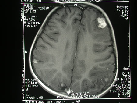
Image c. 3 yr old boy with CNS aspergillosis pt TS. MRI scan pre-amphotericin B
Copyright: n/a
Notes:
A 3 year old boy, quite active and healthy clinically, who has CNS aspergillosis. He was first seen about 4 months ago for a red eye, which turned out to be panophthalmitis; culture yielded Aspergillus spp. He received 2 weeks of iv amphotericin and was sent home by the ophthalmologists. No h/o eye trauma. He returned 2 weeks ago with focal fits, and the MR showed several lesions bilaterally (including ring enhancing lesions) and normal sinuses, and a brain bx showed fungal hyphae (no culture this time). His immune status (normal WCC and neutrophil function so far) was investigated.
He was given conventional amphotericin for 8 weeks, and switched to oral itraconazole. We had to limit the ampho to 0.7 mg/kg owing to toxicity (mainly hypokalaemia).
The MRI scan was repeated at about 6 weeks, and generally showed good improvement (scans e-h). The enhancement/flare were gone but remained in a few lesions, the lesions themselves were all either gone or much smaller. Further investigations revealed the child was immunocompetent.
Patient was switched from amphotericin to oral itraconazole at week 8 essentially on a clinical assessment. Awaiting follow-up.
Images library
-
Title
Legend
-
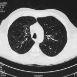
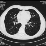
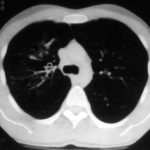
High resolution CT scan images with reconstruction of 1mm thick slices at approximately 10mm increments. The scan shows moderately severe multi-lobar cylindrical and varicose bronchiectasis predominantly centrally and in the upper lungs. There is no mucus plugging seen.
The features are in keeping with allergic bronchopulmonary aspergillosis
 ,
,  ,
, 
-
pt.SB – 6/10/98 – bronchocentric granulomatosis. CT scan showing multiple small nodules of variable size in both lung fields, apparently close to the vascular bundles.

-
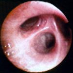
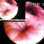
Bronchial oedema.Remarkably oedematous bronchial mucosa, as seen in ABPA.
-
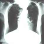
An example of longstanding allergic bronchopulmonary aspergillosis in a patient who has been steroid dependent for over 15 years showing remarkable kyphoscoliosis and honey combing and fibrosis of both lungs.
-
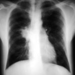
Recurrent pulmonary shadows 1. 6 Jan 1988 – chest radiograph showing right hilar enlargement, consistent with ABPA.
Recurrent pulmonary shadows 1. 3 Feb 1989 – chest radiograph showing right upper-lobe consolidation and contraction consistent with obstruction of RUL bronchus, in ABPA.
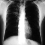
Clearing of pulmonary shadows 3, pt BJ. 5 April 1989 – resolution of shadows seen in February, with a course of corticosteroids.
Recurrence of pulmonary shadows 4, pt BJ. 2 September 1989 – recurrence of pulmonary shadows with an exacerbation of ABPA.
Central bronchiectasis, pt BJ. CT scan of thorax October 1989 showing central bronchiectasis, characteristic of ABPA (and cystic fibrosis).
 ,
,  ,
,  ,
,  ,
, 
-
A typical example of a wet mount of a sputum sample from a patient with allergic bronchopulmonary aspergillosis.


