Date: 26 November 2013
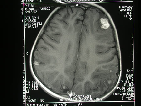
Image c. 3 yr old boy with CNS aspergillosis pt TS. MRI scan pre-amphotericin B
Copyright: n/a
Notes:
A 3 year old boy, quite active and healthy clinically, who has CNS aspergillosis. He was first seen about 4 months ago for a red eye, which turned out to be panophthalmitis; culture yielded Aspergillus spp. He received 2 weeks of iv amphotericin and was sent home by the ophthalmologists. No h/o eye trauma. He returned 2 weeks ago with focal fits, and the MR showed several lesions bilaterally (including ring enhancing lesions) and normal sinuses, and a brain bx showed fungal hyphae (no culture this time). His immune status (normal WCC and neutrophil function so far) was investigated.
He was given conventional amphotericin for 8 weeks, and switched to oral itraconazole. We had to limit the ampho to 0.7 mg/kg owing to toxicity (mainly hypokalaemia).
The MRI scan was repeated at about 6 weeks, and generally showed good improvement (scans e-h). The enhancement/flare were gone but remained in a few lesions, the lesions themselves were all either gone or much smaller. Further investigations revealed the child was immunocompetent.
Patient was switched from amphotericin to oral itraconazole at week 8 essentially on a clinical assessment. Awaiting follow-up.
Images library
-
Title
Legend
-
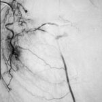
Embolisation 7 – patient WC. Angiogram of the lateral thoracic artery on subtraction film showing grossly abnormal vasculature inferiorly shunting along several anterior intercostal arteries to the internal mammary artery. In addition a pseudoaneurysm is shown.
-
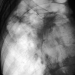
Embolisation 6 – patient WC. Catheter tip in the lateral thoracic artery on screening film.
-
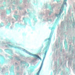
Grocott (silver) stain showing branching septate hyphae fairly typical of Aspergillus in mucus. The apparent right angle branching is unusual.
-
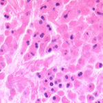
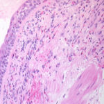
Bronchial mucosa under H & E stain showing numerous eosinophils deep to the mucosa, and mucus in the lumen of the bronchiole.
-
Grocott (silver) stain showing branching septate hyphae fairly typical of Aspergillus in mucus. The apparent right angle branching is unusual.

-
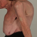
Severe kyphoscoliosis caused by greater than 40 years of prednisolone for ABPA and asthma.

-
These pictures show remarkable curvature of the spine as a result of collapse of the vertebral bodies of the thoracic vertebrae. This is a gross example of steroid-induced osteoporosis. The dose was not large in the last 10 years, typically 5-10mg daily, but multiple high dose courses and slow tapering lead to this outcome.
Her corticosteroid warning card is also demonstrated, as additional steroids are required for any significant illness or surgery, as her adrenal glands had completely atrophied.
Kindly supplied by Prof David Denning, South Manchester University Hospitals NHS Trust, Manchester UK
(© Fungal Research Trust)
 ,
,  ,
,  ,
,  ,
, 

