Date: 26 November 2013
Image c. 3 yr old boy with CNS aspergillosis pt TS. MRI scan pre-amphotericin B
Copyright: n/a
Notes:
A 3 year old boy, quite active and healthy clinically, who has CNS aspergillosis. He was first seen about 4 months ago for a red eye, which turned out to be panophthalmitis; culture yielded Aspergillus spp. He received 2 weeks of iv amphotericin and was sent home by the ophthalmologists. No h/o eye trauma. He returned 2 weeks ago with focal fits, and the MR showed several lesions bilaterally (including ring enhancing lesions) and normal sinuses, and a brain bx showed fungal hyphae (no culture this time). His immune status (normal WCC and neutrophil function so far) was investigated.
He was given conventional amphotericin for 8 weeks, and switched to oral itraconazole. We had to limit the ampho to 0.7 mg/kg owing to toxicity (mainly hypokalaemia).
The MRI scan was repeated at about 6 weeks, and generally showed good improvement (scans e-h). The enhancement/flare were gone but remained in a few lesions, the lesions themselves were all either gone or much smaller. Further investigations revealed the child was immunocompetent.
Patient was switched from amphotericin to oral itraconazole at week 8 essentially on a clinical assessment. Awaiting follow-up.
Images library
-
Title
Legend
-
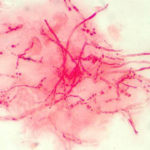
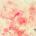
Corneal ulcer – gram stain. Corneal scrapings were taken from a 67 yr old farmer presenting with a corneal ulcer of the right eye. A piece of vegetable matter was embedded in the cornea and scrapings were done. Gram stain (500x magnification) showed numerous septate hyphae. Cultures grew a small amount of A fumigatus.

-
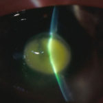
Aspergillus keratitis. Central lesion in aspergillus keratitis following a corneal foreign body which made a good response to topical treatment alone, albeit over 2 months intensive treatment.
-
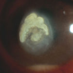
Aspergillus keratitis. B- Severe central aspergillus infection with a “cheesey†looking area of the lesion and hypopyon (fluid level of inflammatory cells in the anterior chamber)
-
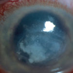
Aspergillus keratitis. A- Severe aspergillus infection with large area of corneal ulceration and deep stromal involvement
-
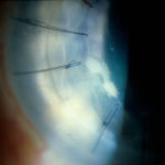
Candida keratitis. Focal candida keratitis as an unusual cause of a suture related infection following corneal transplantation for non infective indication
-
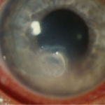
Candida keratitis. Subacute onset of candida keratitis in a young adult in whom dust blew into her eye in Greece. A slightly “feathery†edge to stromal involvement is suggestive of fungal infection
-
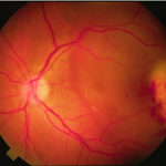
Aspergillus endopthalmitis. Temporal necrosis due to Aspergillus endopthalmitis as part of disseminated disease. No evidence of vitritis. Systemic treatment essential.
