Date: 26 November 2013
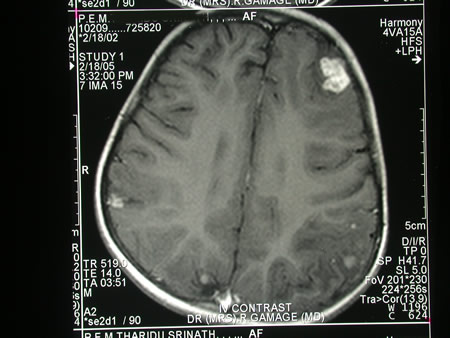
Image c. 3 yr old boy with CNS aspergillosis pt TS. MRI scan pre-amphotericin B
Copyright: n/a
Notes:
A 3 year old boy, quite active and healthy clinically, who has CNS aspergillosis. He was first seen about 4 months ago for a red eye, which turned out to be panophthalmitis; culture yielded Aspergillus spp. He received 2 weeks of iv amphotericin and was sent home by the ophthalmologists. No h/o eye trauma. He returned 2 weeks ago with focal fits, and the MR showed several lesions bilaterally (including ring enhancing lesions) and normal sinuses, and a brain bx showed fungal hyphae (no culture this time). His immune status (normal WCC and neutrophil function so far) was investigated.
He was given conventional amphotericin for 8 weeks, and switched to oral itraconazole. We had to limit the ampho to 0.7 mg/kg owing to toxicity (mainly hypokalaemia).
The MRI scan was repeated at about 6 weeks, and generally showed good improvement (scans e-h). The enhancement/flare were gone but remained in a few lesions, the lesions themselves were all either gone or much smaller. Further investigations revealed the child was immunocompetent.
Patient was switched from amphotericin to oral itraconazole at week 8 essentially on a clinical assessment. Awaiting follow-up.
Images library
-
Title
Legend
-
Nasal aspergillosis in an English Pointer. Photograph of an English Pointer with nasal aspergillosis
Nasal aspergillosis in an English Pointer. Photograph of an English Pointer with nasal aspergillosis

-
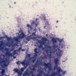
Domestic crossbred cat with disseminated aspergillosis. Diff Quik stained squash preparation of material obtained from thoracotomy of a 3 year old domestic crossbred cat with invasive Aspergillus fumigatus infection. The cat had marked enlargement of the hilar lymph nodes that resulted in a partial tracheal obstruction. This smear was made from portions of the hilar lymph node resected at thoracotomy. Magnification x 132.
-
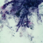
English Pointer with nasal aspergillosis. Diff Quik stained cytological smear of material obtained from the frontal sinus of a 7 year old English Pointer with nasal aspergillosis. This infection was caused by Aspergillus fumigatus. Fungal hyphae are beautifully demonstrated by the Diff Quik stain. Magnification x 200.
-
Nasal aspergillosis in a Schnauzer. Histological section of fungal plaques removed surgically from the frontal sinus of a 5 year old Schnauzer with nasal aspergillosis. This infection was caused by Aspergillus fumigatus. H & E; x 200.

-
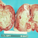
Disseminated aspergillosis in a German Shepherd. Masses of fungal hyphae in the renal pelvis of both kidneys in a young German Shepherd dog with disseminated Aspergillus terreus infection.
-
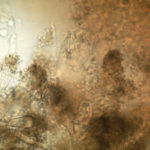
Nasal aspergillosis. KOH preparation of fungal plaques surgically removed from the frontal sinus of a Schnauzer with nasal aspergillosis due to Aspergillus fumigatus.
-
English Pointer with nasal aspergillosis. English Pointer with nasal aspergillosis treated by topical enilconazole injected through surgically inserted indwelling plastic tubes.

-
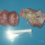
German Shepherd with disseminated aspergillosis. Unilateral pyelonephritis in a German Shepherd dog with disseminated Aspergillus terreus infection.
-
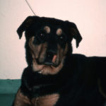
Rottweiler treated with indwelling plastic tubes. Photograph of a Rottweiler crossbred dog treated with indwelling plastic tubes placed surgically into the nasal cavity and frontal sinuses.

-
Rottweiler crossbred dog with nasal aspergillosis. A Rottweiler crossbred dog with nasal aspergillosis due to Aspergillus fumigatus infection. Note the loss of pigment below the nostril on the worst affected side – this finding is suggestive of a diagnosis of chronic nasal aspergillosis in the dog.
