Date: 26 November 2013
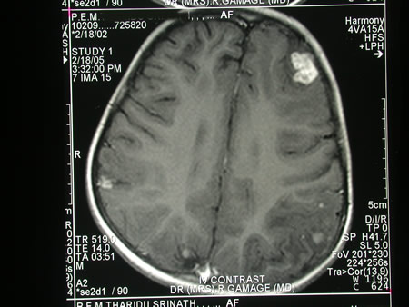
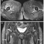
Image c. 3 yr old boy with CNS aspergillosis pt TS. MRI scan pre-amphotericin B
Copyright: n/a
Notes:
A 3 year old boy, quite active and healthy clinically, who has CNS aspergillosis. He was first seen about 4 months ago for a red eye, which turned out to be panophthalmitis; culture yielded Aspergillus spp. He received 2 weeks of iv amphotericin and was sent home by the ophthalmologists. No h/o eye trauma. He returned 2 weeks ago with focal fits, and the MR showed several lesions bilaterally (including ring enhancing lesions) and normal sinuses, and a brain bx showed fungal hyphae (no culture this time). His immune status (normal WCC and neutrophil function so far) was investigated.
He was given conventional amphotericin for 8 weeks, and switched to oral itraconazole. We had to limit the ampho to 0.7 mg/kg owing to toxicity (mainly hypokalaemia).
The MRI scan was repeated at about 6 weeks, and generally showed good improvement (scans e-h). The enhancement/flare were gone but remained in a few lesions, the lesions themselves were all either gone or much smaller. Further investigations revealed the child was immunocompetent.
Patient was switched from amphotericin to oral itraconazole at week 8 essentially on a clinical assessment. Awaiting follow-up.
Images library
-
Title
Legend
-
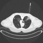
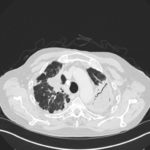
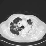
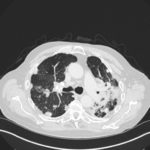
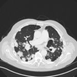
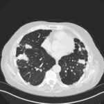
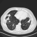
Mr RM is 80 and an ex-coal miner.He developed pneumoconiosis from exposure to coal dust. He also developed rheumatoid arthritis and the combination of this disease and pneumoconiosis is called Caplan’s syndrome.
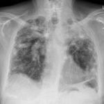
His chest Xray in early 2015 shows extensive bilateral pulmonary shadowing with solid looking nodules superimposed on abnormal lung fields, contraction of his left lung with an elevated diaphragm and a large left upper lobe aspergilloma, displaying a classic air crescent. His CT scan from mid 2014 demonstrates a large aspergilloma in a cavity on the left, with marked pleural thickening around it, which is partially ‘calcified’ towards its base. Inferiorly on other images,remarkable pleural thickening and fibrotic irregular and spiculated nodules are seen, most partially calcified.
 ,
,  ,
,  ,
,  ,
,  ,
,  ,
, 