Description: Selective Plane Illumination Microscopy (SPIM) greatly reduces phototoxicity in comparison with other fluorescent imaging modalities and makes it possible to image living small animals in 3D over extended periods of time. This talk describes an extension of SPIM such that it can be used with specimens on a coverslip rather than in a capillary (inverted SPIM or iSPIM), and a second modification that images the specimen using two perpendicular light sheets (Dual-View iSPIM or diSPIM), resulting in 3D datasets with the same resolution in X, Y, and Z (isotropic resolution)
About the Speaker: Hari Shroff
Hari Shroff is an Investigator at the National Institute of Biomedical Imaging and Bioengineering at the National Institutes of Health. During his post-doc, which was with Eric Betzig at HHMI’s Janelia Farm Research Campus, he focused on the development of PALM (photoactivated localization microscopy) microscopy. Since then he has developed several microscopy techniques, such as diSPIM and instant structured illumination microscopy.
For full tutorial & assessment go to iBiology
Medical and Patient education videos
-
Title
Description
-

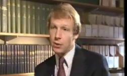
Woodrow Maitland tells of his personal experience of having aspergillosis. Like most people he had never heard of aspergillosis when he was finally diagnosed and treated at the National Aspergillosis Centre, Manchester, UK
-
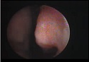
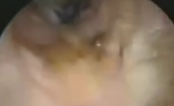
A series of nasal endoscopies – before during and after surgery. Surgical endoscopy by Raphaella Migliavacca. A patient 12 years old with AML undergoes remission following chemotherapy (2 rounds), admitted several months later with high fever and neutropenia. Patient undergoes regime of antifungal therapy including fluconazole, AmpB and voriconazole. (see full case as powerpoint slides presented In Brazil In FocusVII Aug 2009 Raphaella Migliavacca.). Nasal endoscopy was performed both pre-operatively and surgery removed a fungal ball. Biopsy revealed suppurative acute inflammatory findings (middle turbinate) and angioinvasive fungal structures consistent with aspergillosis. Cultures revealed Aspergillus flavus. Eight weeks later another endoscopy post surgery revealed marked improvement and reduced inflammation. The patient was given 12 weeks voriconazole followinf the surgery, followed by 3rd chemotherapy. The patient had recovered and was in complete remission 4 months after the surgical endoscopy.
-
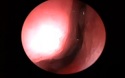
Pre-operative endoscopy by Raphaella Migliavacca.
-
Produced by David Denning and described by Dr Rob Bissett. With kind permission of Gardiner-Caldwell, funded by Janssen-Cilag. Copyright Aspergillus Website.
-
This patient had a small hole in the bone at the base of the skull. He underwent endoscopy through the nose which shows the defect in the skull through which the brain is visible (pulsating). Kindly provided by Hesham Saleh, Consultant Rhinologist, Charing Cross and Royal Brompton Hospital .
-
The Aspergillus Website maintains a collection of clinical videos published on Youtube. Click on the listing at the top to access all videos.
-
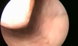
Kindly provided by Hesham Saleh, Consultant Rhinologist, Charing Cross and Royal Brompton Hospital
-

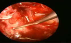
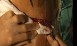
Removing fungal growth from the maxillary sinus via the use of an endoscope.
-
This video shows the removal of the fungal ball through the left nasal passage
-
Produced by Dr Mark Jones and David Denning. Copyright Aspergillus Website.
: Hari Shroff)