Description:
The role of poorly maintained ventilation ductwork in spreading airborne infections is largely ignored because it is ‘out of sight out of mind’, but recent analysis by healthcare professionals confirms that the risks are increasing.
Ghasson Shabha will identify particular threats in healthcare facilities from dirty ductwork. He also points out that fewer than 5% of building air-conditioning systems have been inspected despite regulations making this mandatory. Dr Shabha will look at how planned cleaning strategies can reduce risks and how hospitals can use 3D building information modelling software to identify ‘infection hotspots’.
The healthcare environment can be described as a reservoir for potentially infective agents which can spread unpredictably in a whole array of ways particularly in ventilation and air-conditioning systems making it difficult to effectively control and manage, for those seeking timely information about the patterns of cross-infection. The severity of the problem has been highlighted extensively over the past decade, as part of wider umbrella of what was known as health care acquired infections (HCAIs). Airborne transmission extends over a wide spectrum, and includes many prevalent agents, inter alia Mycobacterium tuberculosis (TB),nosocomial MRSA, Aspergillus fumigatus, Serratia marcescens, norovirus, and other airborne infection.
Dr Shabha was speaking live from Birmingham and this CIBSE ASHRAE Group (cibseashrae.org) webinar was co-sponsored by the B&ES Ventilation Hygiene Group and the Institute of Healthcare Engineering and Estate Management (IHEEM).
CIBSE ASHRAE Group – Unclean, unclean! Health threats from ventilation and air conditioning – 14th Nov 2012 from Tim Dwyer on Vimeo.
Medical and Patient education videos
-
Title
Description
-
Prof. Patricia Muñoz, Division of Clinical Microbiology and Infectious Disease, Hospital General Universitario Gregorio Marañón, Madrid, Spain
-
Dr. Eavan Muldoon, Consultant in Infectious Diseases, University Hospital of South Manchester
-
Prof. Adilia Warris, Principal Investigator, Aberdeen Fungal Group, University of Aberdeen
-
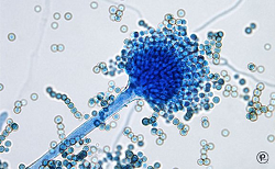
Created by Patrick Hickey for the “Killer Fungus” exhibit at the 2016 Royal Society Summer Science Exhibition
A large dish of nutrient jelly inoculated with several different fungal species and then filmed over 5 days. The different colours of the fungal species used gradually grow to form the image and spell out the message ‘Killer fungus’.
The awareness, diagnosis and treatment of serious fungal infections needs dramatic improvement throughout the world as millions die needlessly every year.
The Royal Society Summer Exhitions are aimed at informing and inspiring the public – children and our future engineers and scientists in particular. Killer fungus is one exhibit amongst many more covering every aspect of science you can think of!
More
http://www.killerfungus.org/ h
ttps://twitter.com/ killerfungus16 https://www.facebook.com/ killerfungus16/ -
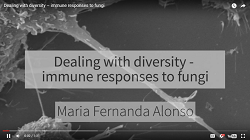
Fungi are everywhere. They’re present in the air, in our food, some even live inside our bodies. But most of us rarely think about diseases caused by fungi. This is because our defences do a really good job in keeping them in check.
However for millions of people whose immune system are defective fungi can cause serious infections that are hard to treat and can be life threatening.
A group of cells called phagocytes play a key role in keeping us safe from fungi. They normally patrol our body so when there’s a breach they are the first to respond. Their function is to seek eat and destroy microbial intruders.
But no all fungi are alike. My Ph.D. project aims to understand how phagocytes tackle such different targets. So far I have found there are huge differences in the rate at which various different fungi are engulfed by phagocytes.
The speed of these processes depends on the chemical composition of the fungi, whether they are alive or dead, and whether they are coated with human proteins that help to mark them as intruders.
Understanding the basic biology behind these processes is the first step towards developing new treatment strategies.
Maria Fernanda Alonso works in Professor Neil Gow’s lab at the University of Aberdeen.
-

Candida albicans is one of the fungal species most commonly causing life threatening infections in vulnerable patients. Our group is studying the mitochondria in Candida albicans cells.
Just like in our cells the mitochondria are the batteries of the cell, producing energy required for growth and in fungal cells can also influence infection.
The mitochondria can influence components of the fungal cell wall and its ability to infect us.
If we understand how mitochondria are influencing these factors, we can develop new anti-fungals or we can use existing anti-fungals in new combinations in order to tackle life threatening infections.
Lucian Duvenage works in Dr Campbell Gourlay’s lab at the University of Kent
-

Every day we inhale hundreds of fungal spores but these in healthy individuals are efficiently eliminated by specialist immune cells called phagocytes which engulf and kill them. However, some human illnesses interfere with this defence mechanism, increasing susceptibility to fungal diseases.
A specialist lung tissue called the epithelium is the first line of contact between the inhaled spores and us, the host. We are working to understand how the lung epithelium interacts with the spores of a common mould called Aspergillus fumigatus.
We have generated fluorescent Aspergillus and combined this with fungal and host specific dyes to directly visulaise this interaction. We have discovered that epithelial cells ingest fungal spores and kill them.
This might provide a critical defence mechanism which is acting while we breathe, and before even phagocytes arrive at the site of the infection.
We are now trying to work out how epithelial cells grab and ingest fungal spores, by using fluorescent fungal mutants and targeted elimination of host proteins.
Once we understand this process in detail we can design new therapies to assist a quicker elimination of the dangerous fungal spores we all inhale on a daily basis.
Dr Margherita Bertuzzi works in Dr Elaine Bignell’s lab at the University of Manchester
-
My research is focused on infections caused by Aspergillus, which is present in the environment all around us. We each inhale hundreds of fungal spores every single day, and for the majority of people this doesn’t cause any problems, but for people whose immune system isn’t working properly Aspergillus can cause serious, even life threatening, infections.
One group of patients who are at particular risk of Aspergillus infection are people with a rare inherited immune deficiency called chronic granulomatous disease, or CGD.
For these patients infection with Aspergillus is frequently fatal, even with appropriate anti-fungal treatment.
My research so far suggests that rather than not mounting a sufficient immune response, the CGD immune system actually over-reacts to Aspergillus, causing significant tissue damage but without clearing the fungal infection.
We’re therefore looking at whether we can use new treatments aimed at reducing inflammation alongside conventional anti-fungal drugs to improve the outcome of these devastating infections for CGD patients.
Dr Jill King works in Professor Adilia Warris’s lab at the University of Aberdeen
-

Cryptococcus, like many fungi, produces spores that are found in the air that we breathe. These spores will be inhaled into our lungs but they do not cause any harm because of our immune defences. However, they can cause life-threatening infections in individuals that have a weakened immune system, for example those that have AIDS or have had an organ transplant.
So, we’re interested in immune cells called macrophages which are needed for immune defence against Cryptococcus.
It is impossible to see how immune cells destroy this fungus during infection because we do not have see-through bodies. So, in order to study this we are using zebrafish. Zebrafish have a similar immune system like our own but are transparent which makes it possible to see how infections happen. Using zebrafish we are testing new ways to enhance immune defences against Cryptococcus.
Ultimately these investigations may lead to development of novel therapies towards Cryptococcal infections.
Alfred Kamuyango works in Professor Simon Johnston’s lab at the University of Sheffield.
-
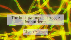
We can view an infection as a battle between the human host and the microbial invader. The outcome of which decides whether the host remains healthy or succumbs to disease. As this battle rages microbial invaders use their hosts as a source of nutrients.
However, the human body has evolved complex systems to limit access to certain essential nutrients in an attempt to starve the invading microbes and prevent disease.
We call these processes nutritional immunity. Therefore, to win the battle and cause disease microbial pathogens must have evolved strategies to thrive in a nutritionally restrictive environment within its infected host.
We are interested in exploring how the human fungal pathogen Candida albicans adapts to limitations to essential trace nutrient zinc. We have identified specific coping mechanisms that are adopted by this fungus in order to deal with nutritional immunity. In response to zinc starvation,Candida albicans dramatically changed their cell shape.
We therefore want to know how this change is regulated and what impact it has on the progression of infection with the ultimate aim to therapeutically manipulate the system and push the balance back in favour of the human host to prevent disease.
Dhara Malavia works in Dr Duncan Wilson’s lab at the University of Aberdeen.