Submitted by HLeSueur on 4 December 2015
Early diagnosis of invasive fungal infections is key to improving survival among immunocompromised patients. However, current diagnostic procedures are limited, particularly with regards to sensitivity and specificity. One area of research which has great potential is the use of radiotracers, which can be used to carry out ‘molecular trapping’, generating intense radioactive signals in target cells. This signal can then be picked up by Positron-Emission Tomography (PET). PET provides excellent sensitivity and resolution and has been an extremely successful clinical imaging technique in other areas, such as oncology. This is due to the specific pharmacokinetics of the molecules utilised for radiotracers (for more information on radiotracers in oncology see article). If an equivalent radiotracer for fungal infections can be found, many patients could benefit from earlier diagnosis and treatment. Due to the prevalence of Aspergillus fumigatus, researchers have recently been trying to develop radiotracers that can target and localise A. fumigatus specifically. However, current methods all demonstrate a lack of active uptake by the pathogen, which means that there is still much research to be done. Huburtus Haas and colleagues at Innsbruck Medical University have therefore been investigating an iron-uptake mechanism as a potential target for a radiotracer (article).

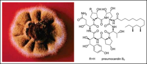
(A) FSC/TAFC is shown in the ferri-form; the ester bonds separating the three N5-acetyl-/N5-anhydromevalonyl-N5-hydroxyornithine residues are shown in green; for TAFC-based nuclear imaging, the iron (shown in red) is replaced by 68Ga. FSC, R = H; TAFC, R = acetyl. (B) TAFC-mediated uptake of iron and gallium into fungal hypha.
Iron plays a vital role as a nutrient for A. fumigatus during infection. The pathogen has therefore developed efficient mechanisms to obtain it from the host (which attempts to commandeer all available iron). One of the methods used by A. fumigatus is known as ‘siderophore-mediated iron acquisition’. A siderophore is a compound produced by A. fumigatus in order to scavange iron from the host. Siderophores can be radiolabelled by replacing the ferric iron (Fe[III]) inside them, with a radionuclide substitute that mimics the original. Huburtus Haas and colleagues have been investigating 68GA, an Fe(III) substitute with a physical half-life of 68 minutes. The substitute compound is misinterpreted as an iron source by the pathogen and therefore accumulates at the infection site. A proof of concept study demonstrated that radiolabelling of siderophores produced by A. fumigatus was easily achievable with 68GA. Furthermore, recent studies in rat models have shown that the radiolabelled siderophore was taken up in infected lung tissue at 20 times the concentration than in healthy lung tissue and was rapidly excreted as an intact complex. This is promising research, though further studies are required to clarify the sensitivity and specificity in a clinical setting.
News archives
-
Title
Date

