Date: 10 February 2014
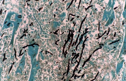
Disseminated, invasive aspergillosis showing dichotomously branching hyphae. Original magnification x150. Stained with Gomori Methenamine Silver (GMS).
Copyright:
Histology kindly provided by Dr Carolyn Burns, Jewish Hospital, Rudd Heart & Lung Center, Louisville, KY. (C) Fungal Research Trust.
Notes: n/a
Images library
-
Title
Legend
-
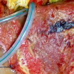
This patient, had had a laparostomy for recurrent intra-abdominal sepsis following on from crohns disease. She was transferred to another intensive care unit and her dressings changed daily. Shortly after, this dark patches appeared on her liver (as seen here A) and her colon. Superficial biopsies and culture showed A.fumigatus invading liver capsule. She responded to amphotericin B therapy.
B shows patient after treatment.
 ,
, 
-
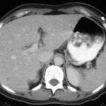
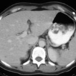
Hepatic aspergillosis, pt KO. Repeat CT scan of the liver showing almost complete resolution of lesions on itraconazole therapy.
-
Image A. The CT scan of her abdomen had the appearances shown here. She also has small pulmonary nodules. Bioposy of the liver revealed hyphae consistent with Aspergillus.
Image B. She responded well to oral itraconazole therapy.
 ,
, 
-
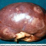
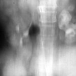
This image shows the pelvis of the left kidney filled with fungal balls. Eventually, after failing amphotericin B therapy, she required a nephrectomy. Her case is reported in Davies SP, Webb WJS, Patou G, Murray WK, Denning DW. Renal aspergilloma – a case illustrating the problems of medical therapy. Nephrol Dial Transplant 1987; 2: 568-572.
-
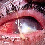
Aspergillus keratitis. Good example of Aspergillus keratitis caused by A.glaucus. Usually A.fumitagus and A.flavus are the causes.

 ,
, 